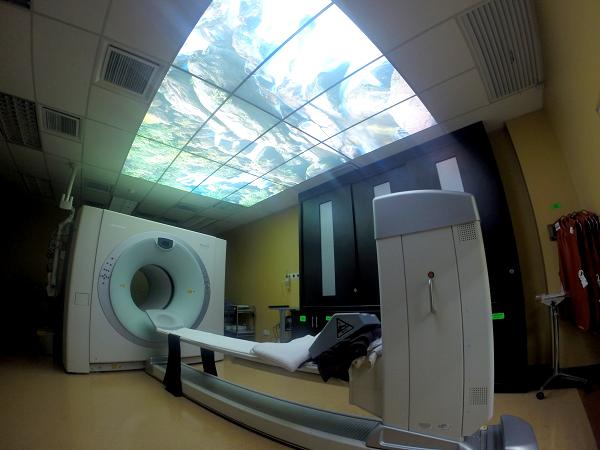
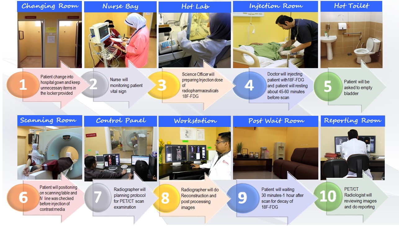
Positron Emission Tomography (PET) machine is the latest imaging modalities in nuclear imaging. It is used to find out the function of cells in abnormal cells especially in cancer. Fluorodeoxyglucose (18F-FDG) is used as radiotracer in PET examination.
The hybrid imaging (combination of PET scan and CT scan) is effective in order to visualize the anatomy and the physiology (function) of the organ at the same time. So the early detection can be done, thus increase the chance for the patient to survive.
PET-CT Examination Procedures
Updated:: 09/04/2018 [muhammadhafiz]
Centre for Diagnostic Nuclear Imaging
Universiti Putra Malaysia
43400 Serdang
Selangor