At PPDN, we actively involve in clinical research looking at specific medical field focusing on our strength in molecular imaging. With the assistance of molecular imaging technique, we are able to help and guide the clinicians in their research.
These are few collaborations that we enganged at the past with clinicians to help them to understand the behaviour of the diseases.
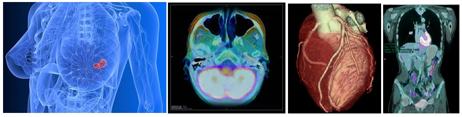
a) Cardiology
Molecular imaging in myocardial viability and perfusion using 18F-FDG and Rb-82 PET/CT. The research was developed with the following objectives:
i) to develop normal LV polar map database for Asian population to be utilized for interpreting viability study
ii) to investigate thesuitable substitute for new protocol for viability study
iii) to highlight the potential of 18F-FDG myocardial viability in identifying patients who will benefit from revascularization. Exploiting FDG PET-CT capability to reduce the number of mis-diagnosed hibernating and infarcted segments
iv) to redefine the normal value of myocardial flow reserve in Malaysian/Asian population
v) to develop normal LV polar map database for Asian population to be utilized for interpreting myocardial perfusion study using 82-Rubidium
b) Oncology
Previous collaboration were with the National Cancer Insitute focussing on the pheochromocytomas, which are also referred to as adrenal paragangliomas. Paragangliomas originate from the neural crest-derived paraganglia of the autonomic nervous system. These tumors originate from catecholamine-producing chromaffin cells. The increased glucose uptake imaged with Flurodeoxyglucose Positron Emission Tomography (FDG PET) is largely dependent on the rate of glycolysis. FDG uptake and trapping occurs because of upregulation of glucose transporters (notably GLUT1 and GLUT3) and HEXOKINASES I and II. The research was conducted using the F-18 FDG PET imaging to allow quantification of glucose uptake on pheochromocytomas patients.
One of the major breakthrough for oncology imaging was the use of F-18 Fluorocholine which were synthesized from our own lab for a small patients group with breast cancer. With the collaboration from clinicians of UPM and Universiti Kebangsaan Malaysia Hospital, we were able to turn the table by proving that F-18 Fluorocholine could be used as a diagnostic marker for patient with breast cancer. The F-18 Fluorocholine is always associated as an excellent diagnostic marker for prostate cancer in PET imaging modality for almost a decade. In some cases, we found those patients who showed negative response to F-18 FDG, showed positive response in F-18 Fluorocholine marker.
c) Neurology
Clinical applications in imaging dementia and vulnerable plagues using 18F-FDG PET/CT. The aims of the study were:
i) to investigate the role of 18F-FDG PET-CT in dementia and vulnerable plague
ii) to develop normal ASIAN database for Statistical Parametric Mapping analysis
iii) to highlight the potential of 18F-FDG in other clinical conditions
iv) to incorporate the18F-FDG into routine clinical practice where indicated
Updated:: 11/04/2018 [muhdhishar]
MEDIA SHARING