Through Malaysia 10th Plan, PPDN is exploring a new niche area which most of the works are focused on translating the fundamental works or laboratory work before it is used to patients.
This newly created wing is called Translational Molecular Imaging Laboratory which covers the niche area of radiopharmaceutical sciences, dosimetry, imaging protocol (PET/CT and MRI), the use of optical fibre as a tool for radiation exposure measurement and genetics. These niche area is best briefly summary as below:-
a) Radiopharmaceutical sciences
In view of increasing significance of radiopharmaceuticals in medical diagnosis, the niche area of cyclotron, radionuclides production and radiopharmaceuticals possesses an important element in nuclear medicine diagnosis. The cliniciansare no longer preferred the conventional ubiquitous radiopharmaceutical F-18 FDG. New radiopharmaceuticals emerge one after another that are able to provide high sensitivity and specificity when approaching a very specific oncology type disease. Hence, the ability to explore new radiopharmaceuticals either from particle accelerator (cyclotron) or generator, F-18 fluorine based or non- F-18 fluorine based is essential in assisting management of nuclear medicine diagnosis.
b) Dosimetry
PET CT is a combined multi-modality imaging that provides anatomical locations and functional imaging. The commonly radionuclide used is 18F-FDG, with a half life of 110 minutes. Radioisotope consists of unstable nucleus. Therefore, it will decay to achieve stable configuration of proton and neutron. During radioactive decay, an unstable nucleus spontaneously decomposes to form a different nucleus, giving off radiation in the form of atomic particles or high energy rays. This decay occurs at a constant predictable rate that is referred to as half-life.
c) Imaging Protocol
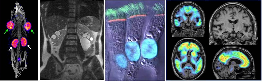
Few studies at PPDN also covers the imaging protocol for PET/CT imaging modality and MRI imaging modality. This is to look for the most efficient imaging protocol that could reduce the burden to patient (dose etc) without compromising the image quality for diagnostic purpose.
One of the study was to look for the effect of contrast-enhanced CT in integrated 18F-FDG PET/CT imaging of cancer patient. The study demonstrated that application of contrast-enhanced CT in PET/CT imaging of cancer patient will cause significant changes on CT values but no significant changes on SUVmax values.
Updated:: 11/04/2018 [muhdhishar]
MEDIA SHARING